
CoCIS_Admin
- Posted on
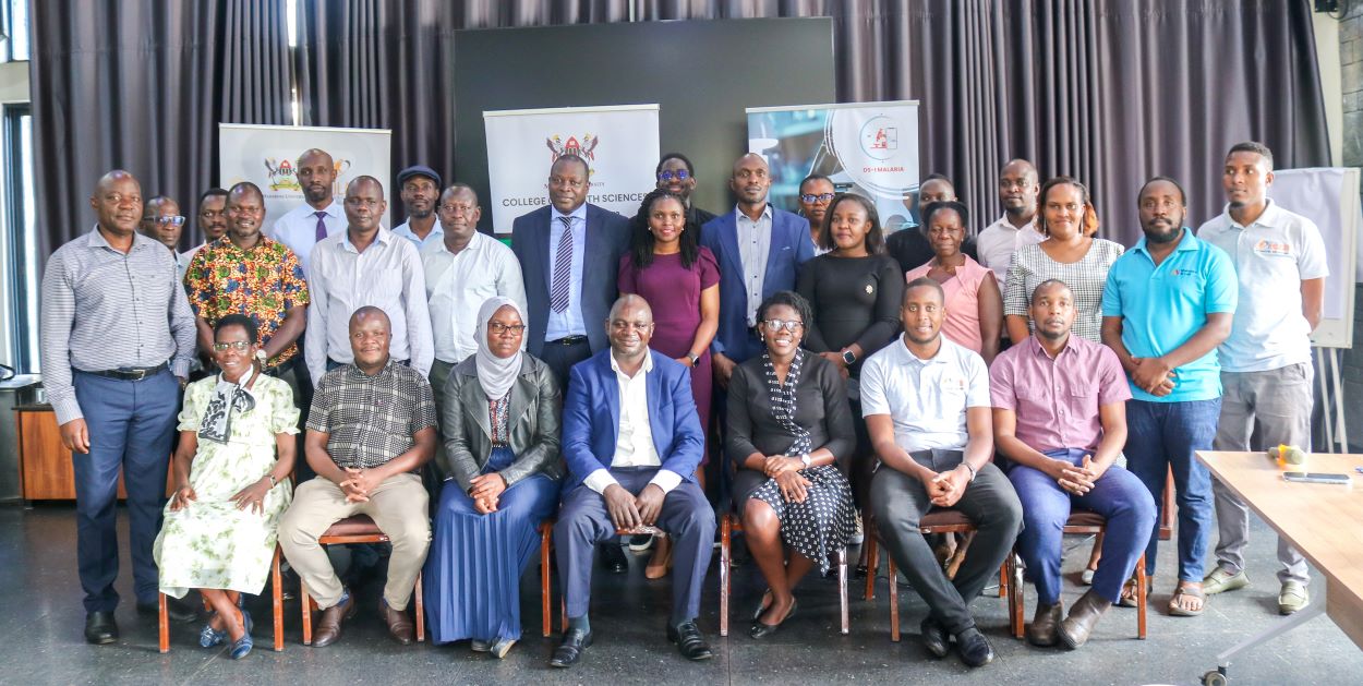
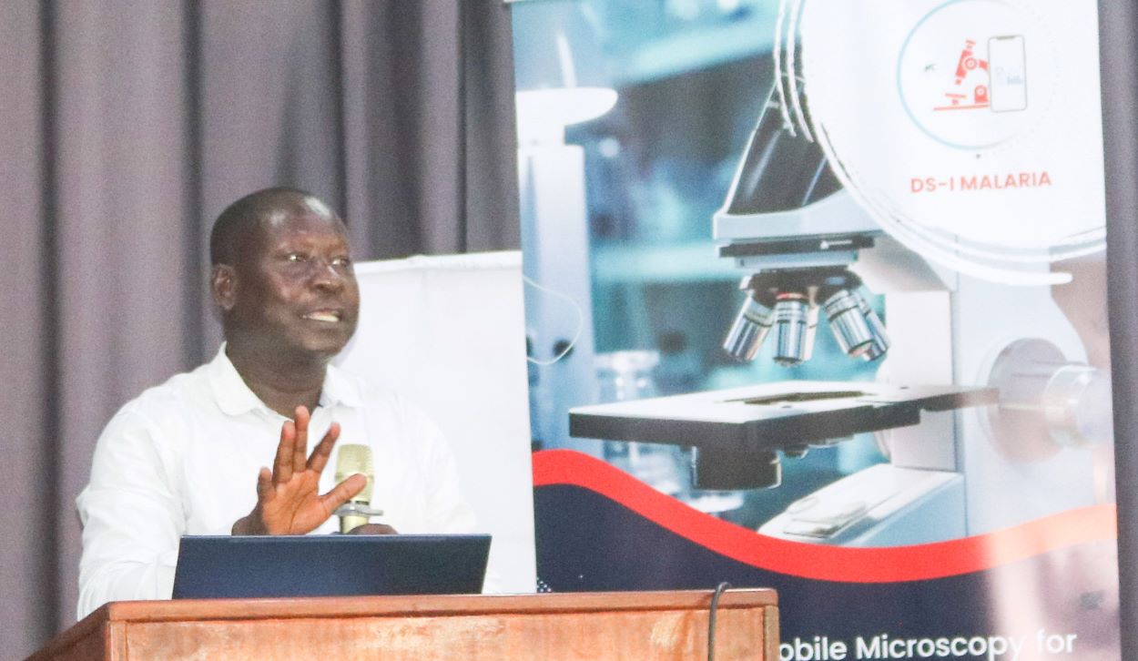
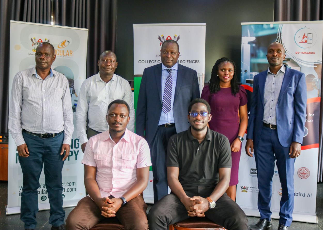
The Automated Mobile Microscopy for Malaria Diagnosis and Surveillance in Uganda (DS-I Malaria Project) has been launched. Field surveys and testimonies from regional hospitals and lower health centres III and IV were also presented during the ceremony held at Fairway hotel in Kampala on 11th March 2024.
The survey team visited the Eastern, Central, Northern and West Nile regions. The primary objective was to see the infrastructure at each hospital and health centres and to see if the lab technicians will accept the application.
DSI Malaria, is a multidisciplinary project that commenced in 2014. This initiative is funded by NIH, a fund that is under the initiative of the Data Science Africa Initiative supporting the work around health and improving health in Africa.
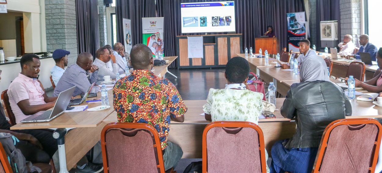
Mak AI Lab in the College of Computing and Information Sciences works collaboratively with the College of Health Sciences, Kiruddu National Referral Hospital, Boston University, Mulago, Sunbird AI, and the Ministry of Health, as well as Okina Project, The project proposition is to leverage Artificial Intelligence for automated microscopy.
The project was officially launched by the Principal, College of Computing and Information Sciences Prof. Tonny Oyana. Oyana said despite all the many control measures, Malaria remains a health and economic burden in sub-Saharan Africa.
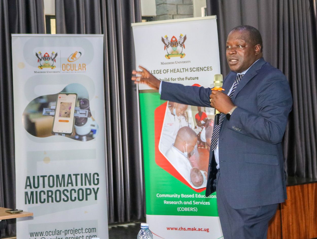
“But malaria is a very important topic of research. Actually, the amount of money that is put into malaria research is very high. Here is a vaccine, but when will it come? Actually, I will be one of the recipients. I would like to go and get one.
Oyana observed that despite Uganda having experienced doctors, one of the biggest challenges is field diagnosis.
“You can go for a malaria test and they tell you have no malaria, but then they leave them. Why don’t we have efficient systems? Since we have suffered and have been vulnerable to malaria for a very long time?
Prof. Oyana describe the research project as very critical
“So the work Dr. Rose Nakasi is doing is to help us diagnose. Microscopy is now being applied to breast cancer, malaria. And the first area was TB. And we hope to expand it so that artificial intelligence can be used in, many areas. that is led by Dr. Rose working with our colleagues at the College of Health Sciences” Oyana said
Project leveraging machine and deep learning techniques and smartphones to improve disease diagnosis. – Dr. Rose Nakasi
Presenting the project overview, Makerere University Principal Investigator Dr Rose Nakasi reported malaria is one of the leading health problems of the developing world whose endemicity has been attributed to poor diagnosis at the lab level which often leads to disease misdiagnosis and drug resistance.
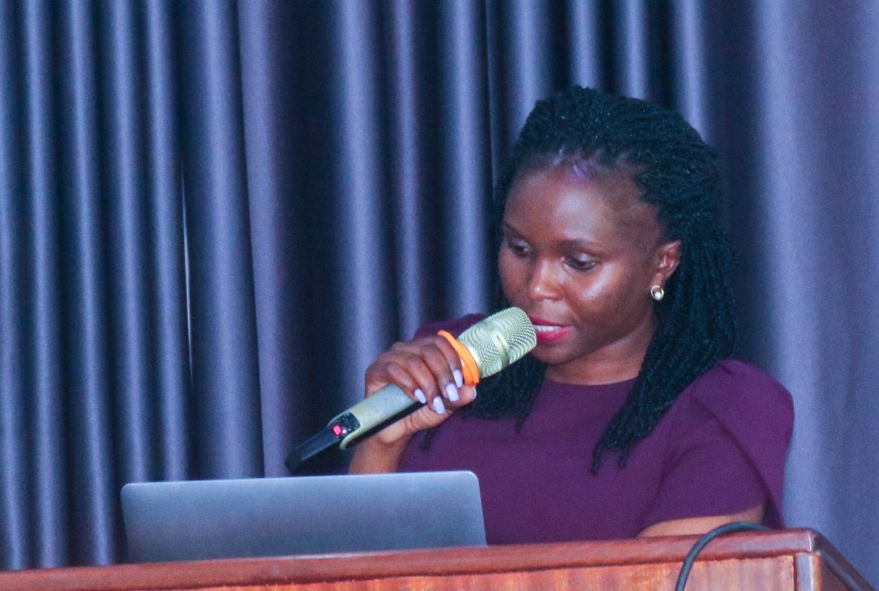
Many developing countries according to Dr. Nakasi are faced with a lack of critical mass of lab technicians to diagnose the disease through a gold standard mechanism of microscopy and this has worsened the already dire situation in some of these Countries.
Nakasi explained that world over, the trending technologies are now based on machine learning and deep learning techniques that can be leveraged with the combination of smartphones to improve disease diagnosis. She however noted that, most of the previous work on automation for microscopy diagnosis has been carried out adhocly in the lab environment and no study seems to give a practical field deployable solution.
“The goal of the proposed research is to develop a rapid, low cost, accurate and simple in-field screening system for microscopy challenges like malaria. Specifically, this study will test and validate developed image analysis models for real time field-based diagnostics and surveillance of malaria.
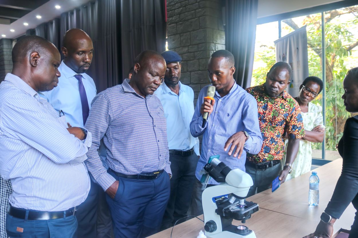
The proposed solution builds from our earlier work on mobile microscopy carried out at Makerere University AI Lab, that has confronted automated microscopy through exploiting recent technological advances in 3D printing to enable development of a low-cost 3D printed adapter. This has enabled attachment of a wide range of Smartphones on a microscope ” Dr. Nakasi said
Furthermore, Nakasi said, the project team has implemented deep learning models for pathogen detection to produce effective hardware and software respectively. The software component is to train machine learning methods to recognize different pathogen objects.
The PI however observed that diagnosis solutions have been ad-hoc in its current state where different conditional settings like image scaling, phone resolutions and grid readings were not standardized and therefore poor performance of the model when deployed in field testing.
She stressed that the infield automated screening trials will therefore involve achieving robust outcomes, through the development of machine learning approaches for standardised field-based microscopy of malaria diagnosis in Uganda and secondly, building a complementary framework for real time surveillance and improved diagnosis of malaria platform through in-field diagnostic studies.
“The point-of-care field-based diagnostic system proposed here addresses a major unmet public health malaria screening and surveillance need to reliably inform public health interventions in malaria control and prevention “the PI affirmed.
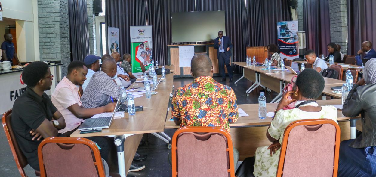
Project team’s surveys health facilities to understand the status of malaria and microscopy in Uganda: Fewer lab technicians and infrastructural challenges reported
Presenting the field survey and testimonies, Dr. Andama Alfred from Mulago National Referral Hospital said, a survey was conducted in four regional blocks encompassing a hospital and health centers III and IV to articulate the gaps that the project wants to fill using the application and also to have a buy in of lab technicians who are the users of the microscopes.
The team according to Dr. Andama met hospital directors and health centre managers in the Eastern, Northern, Central and West Nile regions to understand how the ministry of health is structured, and the health system in terms of care and service delivery
“Each group moved with a set of clear checklist to tease out information that could be useful to leverage this project. The primary objective was to see the kind of infrastructure at each hospital and health centre and also be able to see if lab technicians will be able to accept this applications routine if this project is to be implemented” Andama explained.
Andama reported that overall, the team had good reception with some challenges but the data showed the highest prevalence was in 2009 in West Nile where the equivalence was about 33 but because they went to selected areas, most dispatches of RTD were positive.
Malaria areas reported 70-90 positivity while another interesting outcome of the survey was that sometimes lab technicians run out of these RDTs and report to microscopy.
“And still, under microscopy we found slides reported positive in the book and outside the lab we found children and mothers lying with high fever waiting for results.
From the check list we started with the number of lab technicians many of them in the lower facilities lacked the smart phones and were non-committal to use the smart phone.”Andam reported.
Speaking on the relevant things leveraging the study, Dr. Andama reported that every lab had a microscope, majority with different versions of the Olympus type and majorly did free staining and B staining of Malaria and Tuberculosis.
Andama also reported that there were issues to do with space, running water and workload with most labs run by one person assisted by an intern as the second one who report RTD malaria basing on the same standard.
“We learnt that malaria seems to be a bigger problem than what we see in Kampala. The gap in personnel is real. After lunch, some lab technicians don’t come back as they have to examine all other specimens leave alone malaria. We need to dig deeper into the issue of over diagnosis of malaria. Is it a technical, clinical or stain issue?” Andama wondered.
He reported that the assumption people had in the labs was that once AI is introduced, it will give them time but suggested to go beyond malaria to automate the deposit examinations such as urine.
Andama also reported that in central region, health managers had computers were donated by projects such as IDI not allowed for annotating images while the gene experts have laptops but their labs are a no- go- zone for fear of viruses.
Research team commended and challenged to guide the whole world
Speaking on field experience in malaria research and what researchers should know, Dr. Adoke Yeka commended the research team imploring them to do good work to guide Ugandans and the whole world about this diagnostic device noting that it is more sensitive and time saving .
“The program welcomes such key innovations and research areas. You are moving along the right direction. The quest for quality accurate testing of malaria using AI is a game changer to improve diagnostics in real time”. Dr. Yeka commended
Dr. Yeka noted that Malaria is a big problem affecting all people with pregnant women and children being more vulnerable due to low immunity, and is a biggest cause of sickness and ill health and death in Uganda.
Yeka said , one of the key strategies to control malaria is accurate diagnosis and prompt treatment of the cases.
“We have a lot of challenges when it comes to diagnosis since the disease affects so many people. You need to have the capacity to diagnose so many people who have features suggestive of malaria and tools in place”, he said
He indicated that the gold standard for diagnosing malaria is microscopy in most health facilities in Uganda. He noted that microscopy requires the right microscopes, well trained people with the right skills because results depends on the reader of the equipment.
He emphasized that although there have been recent developments in malaria diagnosis, they come with challenges of relying or detecting antigens in the blood. He observed that even if the pathogens are dead, these rapid tests continue detecting leading to over diagnosis of malaria.
He also noted that most of the new test kits are limited by conditions as they need to be stored under specific conditions.
AI for social good
Dr. Ernest Mwebaze from the Sunbird AI underscored the role of AI in helping to leapfrog from traditional infrastructure. He said, with limited medical personnel and infrastructure challenges, AI can do a lot to move the health sector and improve service delivery.
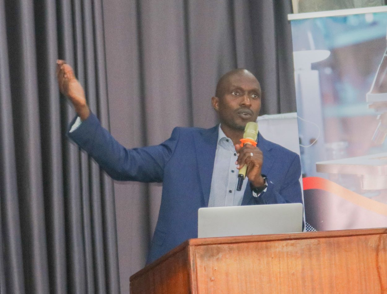
“Social good is about people coming together to do something good for society. There are many areas of social good where AI can help. Social good is about learning patterns. AI is to mimick the intelligent patterns of humans and putting them in a machine normally related to five senses”, Dr. Mwebase explained.
“If given several patterns of a phenomenon, for example several images of slides of malaria that are representative of a phenomenon, AI can learn. Humans have intelligence and AI can amplify human intelligence and make it artificial” he added
As computer scientists, Dr. Mwebaze said, the biggest contribution they can make is looking at how computer science can have a significant good.
He said they have been visiting health centers in Uganda where some labs are busy getting over 200 tests for malaria testing in a day with less than three lab technicians.
“If you automated this process you will be saving time and making the system more accurate. The labs are small with too many patients exposed to diseases. AI is the right system and solution needed”, he recommended
At Makerere University, Mwebaze said, AI has been used to diagnose disease crops in agricultural fields, environmental monitoring of the air quality and pollution.
He said AI uses image data, and could be any sort of data, for example, monitoring TB by listening to TB sounds. From representation, experts can learn some sort of model on top of representation that relate the image to classification and detecting where parasites are.
Read more about the project milestone attached as recorded from the PI Dr. Rose Nakasi.
By. Jane Anyango
Communication Officer